Medical Ultrasound Imaging
Saturday, 27 April 2024
Info Sheets Out- side     | 'Dynamic Range' Searchterm 'Dynamic Range' found in 15 articles 1 term [ • ] - 4 definitions [• ] - 10 booleans [• ]Result Pages : • • 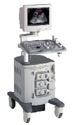 From ALOKA Co., Ltd.;
'A Platform for Digital, Pure-Beam Imaging The high-performance, ALOKA ProSound SSD-3500 utilizes advanced ProSound technologies including: Fully digital beam former A wide dynamic range, 12-bit A/D converter Multi beam processing. The SSD-3500 also helps you achieve more efficient examinations. Its ergonomic, user-friendly design enables you to customize the system according to your specific application needs.'
Device Information and Specification
APPLICATIONS
CONFIGURATION
Compact, portable, dual dynamic display
Color Flow, Power Flow, Spectral Doppler, Real-time Free Angular M-Mode, Tissue Harmonic Imaging, Quint Frequency Imaging, Pure Harmonic Detection
STORAGE, CONNECTIVITY, OS
Data Management Subsystem (iDMS), DICOM-Worklist
DATA PROCESSING
12-bit analog to digital converter
•
(CAI) Color amplitude imaging shows the amplitude of the Doppler signal from moving blood flow. CAI is an ultrasound technique with increased dynamic range and flow sensitivity. The sensitivity of Doppler ultrasound increases markedly in conjunction with the use of vascular contrast agents. See also Amplitude Map, Amplitude Indicator. •
Process of conversion of continuous (analog) signals, such as the detected ultrasound or MRI signal (voltage), into numbers. This is carried out with an analog to digital converter. There are two kinds of discretization involved: the voltage is only measured (sampled) at particular discrete times and only voltages within a particular range and separated by a particular minimum amount can be distinguished. Voltages beyond this range are said to exceed the dynamic range of the digitizer. •
QB-mode (Quadratic Brightness-mode) images are gray scale images from the quadratic component. QB-mode achieves higher contrast and increased dynamic range than the standard B-mode ultrasound images, without loss in spatial resolution.
Further Reading: Basics:
Result Pages : | Share This Page Look Ups |
Medical-Ultrasound-Imaging.com
former US-TIP.com
Member of SoftWays' Medical Imaging Group - MR-TIP • Radiology TIP • Medical-Ultrasound-Imaging
Copyright © 2008 - 2024 SoftWays. All rights reserved.
Terms of Use | Privacy Policy | Advertise With Us
former US-TIP.com
Member of SoftWays' Medical Imaging Group - MR-TIP • Radiology TIP • Medical-Ultrasound-Imaging
Copyright © 2008 - 2024 SoftWays. All rights reserved.
Terms of Use | Privacy Policy | Advertise With Us
[last update: 2023-11-06 01:42:00]